As an industry expert, Ms. Howard has unique access to just about any aesthetic technology. She elected to pursue the mole removal through the use of PLASMA FIBROBLAST due to her confident belief in its ability to safely remove skin growths while managing the remaining skin to not leave any scars. "My mole rested in a tricky/sensitive area (sternum) where excisions have a greater risk of leaving scar. While older methods like freezing or cutting it would most likely have left to scar, I learned about the science of the Plasma Fibroblast technique and was convinced that this was the way to go."
Upon deeper review on her entire journey, Ms. Howard explains that her provider performed a special technique with the 'plasma pen' to extract the tissue mass and also 'build up' the skin around the mole area so it would heal flat- and not leave a concave situation that most mole removals tend to leave behind.
"I have been hesitating to get rid of this mole for many years because every doctor I spoke to talked me out of it... and was firm on the fact that there was no way for this to be removed without a gnarly scar. Then one day, I met with Mario Barton (CEO of
Nuvissa) and Rachel Levy (trainer)- who both gave me tremendous education in the technology. This all gave me peace of mind alongside the promise that the aftermath was going to be absolutely favorable. Sure enough, weeks later (see image enclosed), the plasma fibroblast did not disappoint. No scar!"
DIAGNOSTIC IMAGING UNDER THE SKIN
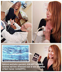
10/21/2022- Ms. January Howard (Ft. Lauderdale, Fla), national expert trainer in minimally invasive facial enhancement procedures (Chemical Peels, PRP, Micro-needling, Biologic fillers) meets renowned medical diagnostic imaging specialist
Dr. Robert L. Bard in his NYC office to discuss a collaborative educational program about image guided procedures for medispas and aesthetics professionals. During this meeting of the minds, they confirmed the need for pre and post-op monitoring and the power of ultrasound technology as the future of safety techniques for the growing list of clinical aesthetic enhancement protocols.
 |
| Scanning a mole with the Doppler ultrasound |
Deeper insight came to light about the technology arose from a real-time diagnostic scenario when Dr. Bard was conducting a standard demo of his scanning equipment directly on Ms. Howard. Dr. Bard noticed a curious raised 7mm mole on her sternum and offered to screen it- partly for her peace of mind. “I have been concerned about that mole for years alongside 30 others all over my body”, stated Ms. Howard. “To be at the right place with Dr. Bard (a cancer imaging expert) is such a blessing… he toured me through what’s under and on my skin in a way that no clinician had ever done before. His real-time scanning and his extensive knowledge in reading anomalies is truly a gift to the aesthetics community!”
Thanks to the 3D Doppler (blood flow) scan of the mole, Ms. Howard was relieved to receive an official medical report that this growth was indeed benign, and ready for a future excision. Dr. Bard’s assessment was just one example of the diagnostic power of real-time scanning—a vital resource for any aesthetician working on or under the skin. “To identify potential land mines for occlusion offers a major advantage in ‘lawsuit prevention’ for any practitioner… with today’s high powered probes and functionalities like elastography and blood flow readings, gathering biometric data is made readily available for just about any budget.”
January Howard is the CEO of MedSpa 101 (
medspa101.com), an aesthetic practice consultancy group and expert trainer in specialized clinical modalities. She is a chapter co-author in Dr. Bard’s upcoming Springer textbook “INDUSTRY REVIEW OF THE AESTHETIC INDUSTRY” along with some of the top names in the aesthetics community- to launch this spring of 2023. Additional information on this textbook is available at:
www.dermalscannyc.com
TRAUMA AND TOXINS: Non invasive diagnostics
Written by: Robert Bard MD
The human body is continually assaulted by harmful forces which may be obvious (trauma and burns) or subtle and dismissed as the “flu or nerves” (chronic poisoning and delayed hidden scarring). However, in the unregulated world of fillers, patients and physicians often discover unexpected findings (forgotten surgical sutures) and complication as potential medicolegal traps. One picture is worth a thousand words and one image may launch a thousand lawsuits while possibly giving birth to a new medical image guided treatment paradigm.
Soft tissue trauma causes a black and blue area but subcutaneous pathology is best imaged by ultrasound. The normal dermal layer is light gray on scans while inflammation is dark gray and fluid (blood) is black. Dermal ultrasound has been used for 30+ years to find skin cancer and guide scar treatment so the use in subacute trauma victims is a logical progression of this portable and non invasive technology. Foreign bodies such as glass and splinters are easily visible as bright white reflectors that directs the surgeon to the exact removal site under ultrasound guidance with minimal tissue “exploration” Fillers have characteristic echo pattern where HA products appear as black globules when they coalesce. Often the HA injected aliquot disperses immediately leaving a diffuse hazy picture. Complications of fillers are well described in recent textbooks. A special case is free silicone having specific “snowstorm” pattern that is commonly seen in breast imaging of ruptured implants. The theoretical possibility of immune system compromise by free silicone is still being studied.
FIBROTIC SCARRING
Elastography shows scar tissue quantitatively in the liver parenchyma but also in traumatized muscles and tendons. The “elastic” properties of tissue are used worldwide for cancer diagnostics because malignant tumors are rock hard and “gritty” with the needle biopsy while benign lumps are soft. Ultrasound maps tissue signatures of free silicone has a mean gray (MG) value 35-40 on a black to white scale.
SKIN OF COLOR
The headline from the 2022 fall issue of PSORIASIS ADVANCES noted inflammatory disease is often misdiagnosed in skin of color. Bruises, burns and infections are detected by color-blind ultrasound as dark areas in the light gray tissues often highlighted by a “ring of fire” blood flow reaction to the local tissue reaction
(Fig 1-L) 45 year female with collagen disease chief complaint of fullness post filler. Sonogram at 18MHx shows a 0.2mm epidermis which expands to 9mm over the tender area. Black HA filler caps the 16x16mm light grey focus of free silicone. Under ultrasound guidance the needle depth was recalculated to avoid injecting and possibly dispersing the silicone material.
REFERENCES
2014 Bard/Marmur Mt Sinai Clinical Trial on Hyaluronidase Efficacy
2020 Bard R: Image Guided Management of Covid Lung Disease 2020 SpringerBerlin
2021 WTC Occupational Cancer Registry: Statistics from Ground Zero 2001
2021 International Inflammatory Disease Symposium NY Academy of Medicine
2022 National Psoriasis Foundation 20:31 NPF Advances
2023 Bard R: Proceedings; Therapeutic Ultrasound Committee of American Institute of Ultrasound in Medicine
REFLECTANCE CONFOCAL MICROSCOPY-
THE LATEST IMAGING ADVANCEMENT FOR DERMATOLOGISTS
Feature updated from original publishing (8/19/2019)- The modern era of diagnostic clinical imaging continues to expand in areas of optimal speed, sensitivity and feasibility as part of its continued pursuits to bring a non-invasive diagnostic modalities to our treatment community. The Reflectance Confocal Microscopy (RCM) gives dermatologists a major upgrade (over age-old microscopy) in their ability to assess pathologic and physiologic conditions of the skin with a higher level of clinical accuracy, greatly supporting the reduction of calls for biopsies of benign lesions. Responding to the limitations of biopsies and conventional screening methods, the non-invasive movement brings a heightened level of performance and responsiveness in areas of resolution, magnification, depth, contrast, and immediate results from bedside. Please see Dr. Manu Jain's complete technology tour
@ Modern Diagnostic Science


No comments:
Post a Comment